A Nashik hospital cannot approve a Varex panel from a model name, nominal size, or wireless claim alone. You need to verify the exact detector configuration, clinical specifications, Indian supply and service terms, and its compatibility with the generator, workstation, PACS, and printer before issuing a purchase order.
Key takeaways
- Name the exact Varex model, variant, and complete acquisition subsystem.
- Request the current datasheet, regulatory documents, warranty terms, and service escalation path.
- Test image transfer, DICOM workflow, battery operation, and X-ray-room compatibility before acceptance.
- Compare panels by handling, durability, workflow fit, support, and total ownership effort.
Which Varex detector configuration is actually being quoted?
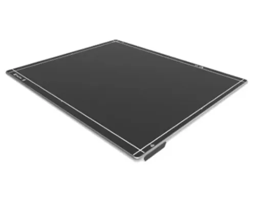
The purchase order must name the exact Varex detector model, variant, and complete acquisition subsystem—not just “Varex flat panel.” For varex detector india details for hospital radiology buyers in nashik, request the current datasheet revision, Indian importer or authorised channel, regulatory documents, warranty exclusions, and Nashik service escalation path.
| Model | Quoted reference to compare | Fit and verification |
|---|---|---|
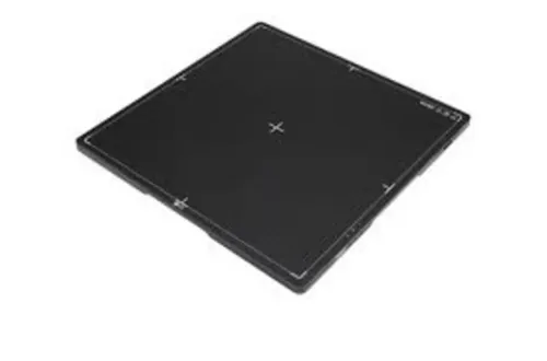
| PaxScan 4343R | Approximately 43 × 43 cm, 139 µm pixels, 3,072 × 3,072 matrix | Full 17 × 17-inch-class coverage; confirm active area, R/RF or wireless variant, tray fit, and collimator field |
| PaxScan 4336R | Approximately 43 × 36 cm, 139 µm pixels, 3,072 × 2,560 matrix | Check chest bucky, table, cassette-tray, and retrofit geometry; compare active area, not nominal size |
| Other quoted configuration | Model-specific dimensions, matrix, options, and ratings | Reject family-level descriptions; require the exact part number and India supply confirmation |
Your varex detector specifications india comparison should require these items in writing:
- Detector technology and scintillator, pixel pitch, matrix, bit depth, spatial resolution, weight, battery type, runtime, charging time, wireless standard, wired backup, load rating, protection and temperature ratings.
- Automatic exposure detection, waterproofing, and impact or drop rating, because the model name alone proves none of them.
- Workstation, generator interface, processing software, DICOM services, PACS/RIS connection, charging, calibration, software updates, and service responsibility.
- Post-replacement gain and offset calibration, bad-pixel-map verification, exposure-index checks, orientation checks, and a signed constancy baseline.
Which specifications affect clinical work and detector handling?
Pixel pitch is only a sampling measure, not a promise of diagnostic detail. For a varex dr panel for hospitals, compare DQE, MTF, noise, lag, ghosting, usable exposure range, and processing settings under the same RQA beam qualities, using IEC 62220-1-1 results where available.
A smaller pixel can still perform worse if noise rises or detail is softened by processing.
Detector construction also changes dose selection and handling:
| Option | Clinical and handling implication | Evidence to request |
|---|---|---|
| CsI scintillator | Usually offers better dose efficiency at comparable sampling; useful for routine and portable imaging | Measured DQE and MTF curves, test beam conditions |
| GOS scintillator | Can bring different cost, ruggedness, and application trade-offs | Scintillator type, construction, and equivalent test data |
| High bit depth | Preserves more signal range, but does not repair poor exposure or processing | Bit depth, exposure range, and exposure-index behaviour |
Require the signed specification sheet to state the active imaging area, detector dimensions, pixel pitch, matrix size, spatial resolution, weight, maximum patient load, battery type, runtime, charging time, wireless standard, and wired backup. Confirm whether automatic exposure detection, waterproofing, and mechanical retention are included options rather than assumed features.
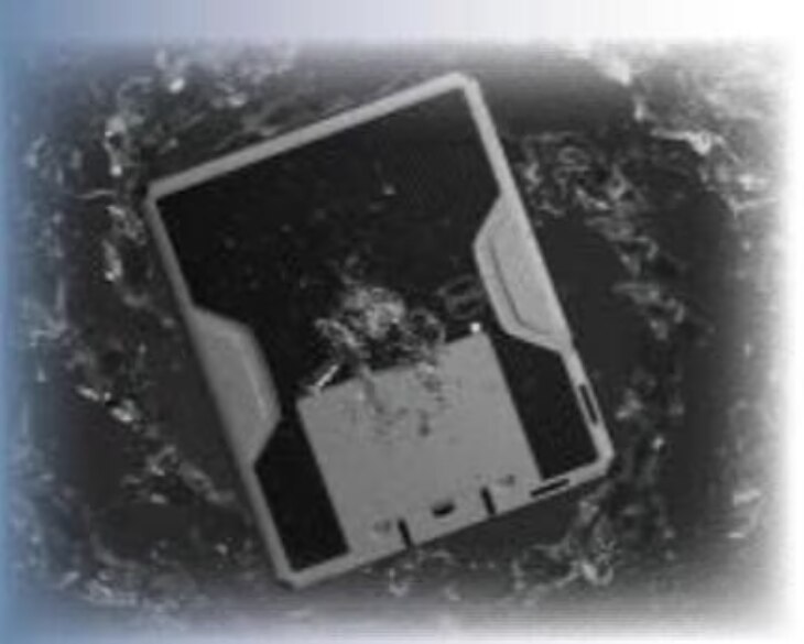
For trauma and bedside work, weight, grip, battery recovery, AED synchronisation, and image orientation matter as much as resolution. Check the ingress-protection rating, operating and storage temperatures, drop or impact rating, and protection against fluid or disinfectant damage.
After a panel swap, demand gain and offset calibration, bad-pixel-map verification, exposure-index checks, orientation checks, and a recorded constancy-test baseline.
Will the panel work with your X-ray room, portable unit, and DICOM workflow?
A matching cassette size does not prove compatibility. For a Varex DR panel for hospitals, test the active area against the bucky, wall stand, and table tray, then confirm generator triggering, exposure synchronisation, automatic exposure detection, detector communication, mechanical retention, and image orientation.
A portable unit needs the same checks, plus reliable wired or wireless connection during bedside exposures.
Require the quotation to identify:
- Generator interface, supported exposure modes, bucky and wall-stand integration, table compatibility, and portable-system compatibility
- Acquisition workstation, image-processing software, detector calibration tools, charging equipment, and wired backup
- RIS/PACS connection, DICOM Modality Worklist, Modality Performed Procedure Step, Storage, and Query/Retrieve
- DICOM Conformance Statement for Basic Grayscale Print Management, because the panel itself does not provide DICOM printing
- Image storage destination, retention responsibility, routing rules, and print-server or DICOM printer requirements
- Responsibility for installation, calibration, software configuration, and post-replacement testing
During commissioning, send a worklist study through the complete path: patient selection, exposure, review, PACS storage, retrieval, and print output. Check patient name, accession number, laterality, pixel spacing, and orientation in the DICOM header; one phantom image will not expose a failed worklist match or incorrect routing.
For a hospital DR detector in Nashik, place gain and offset calibration, bad-pixel verification, exposure-index checks, and a constancy-test baseline in the service scope.
What must Nashik buyers verify about wireless operation, supply, and service?
A wireless quote is incomplete until it defines the detector subsystem, not just the panel. Require separate line items for the acquisition workstation, generator interface, image-processing software, DICOM services, PACS/RIS connection, charging equipment, calibration, warranty, and service responsibility. A buyer searching for Varex detector India in Nashik needs the exact model and variant in writing.
1. Confirm detector pairing, approved access points, network standard, authentication, encryption, VLAN requirements, and wired backup. Ask what the workstation displays when wireless communication fails, whether exposures are stored locally, and how queued images are recovered without duplication.
2. Record battery chemistry, charging method, charge time, runtime at a stated workload, spare-battery price, and charging-cycle warranty. A “wireless” label does not prove readiness for continuous emergency-department use.
3. Identify the manufacturer, Indian importer or authorised channel, and Nashik service provider separately. Before ordering, obtain current Indian regulatory documents, importer details, exact-model availability, promised lead time, warranty exclusions, and the escalation route.
4. Put replacement-panel support in the contract. A panel swap requires gain and offset calibration, bad-pixel-map checks, exposure-index verification, orientation checks, and a new constancy baseline; it is not a plug-in repair.
5. Ask who supplies replacement batteries and parts, delivers software and security updates, and responds on site in Nashik, with response and resolution times stated.
Spark Medical Services can add value by documenting this complete system scope and coordinating installation, calibration, and support for a hospital DR detector in Nashik.
Related product
 | DR System ₹950000 The LUMEN HD Detector by Varex Imaging represents a major advancement in digital radiography technology. View product → |
How should a hospital accept and compare the installed detector?
Accept the panel only after testing the complete detector subsystem, not just one phantom exposure. Record the quoted model, serial number, software version, calibration date, and constancy-test baseline.
Use the same generator, RQA beam quality, exposure settings, phantom, processing profile, and display for every comparison; request DQE and MTF evidence measured under IEC 62220-1-1 where available.
- Check spatial resolution with a line-pair or bar-pattern phantom, then inspect uniformity, bad pixels, lag, ghosting, noise, and visible artifacts at low and routine exposures.
- Repeat exposures to confirm reproducibility. Record exposure index against the console’s target range; a plausible-looking image with a drifting or falsely reported index is a dose-management failure.
- Verify gain and offset calibration, bad-pixel-map application, exposure synchronisation or AED response, detector orientation, and image rotation after installation or panel replacement.
- Send worklist orders from RIS to the workstation and confirm patient ID, accession number, study description, laterality, pixel spacing, rows, columns, and photometric interpretation in the DICOM header.
- Route images to PACS, retrieve them, and print them if printing is part of the workflow. Check that printed orientation, laterality, annotations, grayscale, and scale match the approved image.
| Test | Acceptance evidence | Comparison point |
|---|---|---|
| Spatial performance | Resolution, uniformity, DQE and MTF records | Same beam quality and processing |
| Workflow | Correct headers, worklist, PACS and print output | No manual retyping or routing failure |
| Service readiness | Calibration record and baseline signed | Replacement and future constancy testing |
These records are the practical Varex detector India details for hospital radiology buyers in Nashik and distinguish Varex detector India in Nashik from a quotation that proves only physical installation.
Frequently asked questions
Which Varex detector configuration should a Nashik hospital quote?
Name the exact Varex detector model, hardware variant, acquisition software, accessories, and complete acquisition subsystem in the purchase order. Attach the current datasheet revision so the delivered panel can be checked against the quoted configuration.
Which Varex detector specifications affect clinical work and handling?
Compare active imaging area, pixel pitch, image matrix, image acquisition time, detector dimensions, weight, ingress protection, impact resistance, battery arrangement, charging method, and warranty exclusions. These specifications affect positioning, portability, cleaning, repeat imaging, and staff workload.
Will a Varex DR panel work with an existing X-ray room or portable unit?
Confirm the panel’s generator interface, exposure synchronisation method, software and driver requirements, wireless network needs, mounting dimensions, and supported DICOM services. Test the detector with the intended fixed room and portable X-ray unit rather than relying on a catalogue match.
What should Nashik buyers verify about wireless operation and service?
Ask for wireless frequency and network requirements, battery runtime test conditions, charging equipment, encryption details, replacement-battery terms, Indian importer or authorised-channel records, warranty exclusions, spare-part availability, and the Nashik escalation process.
How should a hospital accept and compare an installed Varex detector?
Record the delivered model and serial number, inspect the panel and accessories, test exposure triggering, image transfer, DICOM worklist and storage, battery charging, detector calibration, and image quality. Compare results with the purchase specification and retain signed acceptance records.
Keywords